Diffuse unilateral subacute neuroretinitis
Diffuse unilateral subacute neuroretinitis (DUSN) is a rare condition that occurs in otherwise healthy, often young patients and is due to the presence of a subretinal nematode.[2]
| Diffuse unilateral subacute neuroretinitis | |
|---|---|
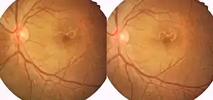 | |
| Left eye retina photograph shows moving nematode larva at the macular area [1] |
Signs and symptoms
The clinical findings in this disease can be divided into acute and end-stage manifestations:
In the acute phase, patients often present with decreased visual acuity, vitritis, papillitis, and crops of gray-white or yellow-white outer retinal lesions. The clustering of the retinal lesions is important because this often helps to localize the causative nematode.
If left untreated, patients ultimately develop late sequel, which may include optic atrophy, retinal arterial narrowing, diffuse retinal pigment epithelial changes, and an abnormal electroretinogram. The late findings of this condition are often misinterpreted as unilateral retinitis pigmentosa.
Cause
DUSN may be caused by a helminthic infection with Toxocara canis, Baylisascaris procyonis, or Ancylostoma caninum. The characteristic lesions are believed to result from a single nematode migrating within the subretinal space. Although previously thought to be endemic in some areas, that belief was likely due to under awareness. DUSN has been diagnosed in patients in many countries and climates including America, Brazil, China and India.[1]
Treatment
If the nematode can be seen by an ophthalmologist, which occurs in less than half of cases, it should be treated with photocoagulation for extramacular location and surgical removal in case the larva is lying in the macula. After the worm is killed, visual acuity loss usually does not progress. Alternatively, Antihelminthic treatment such as high dose oral Albendazole and prednisolone may be used.[1]
Laser photocoagulation is the treatment of choice in cases of DUSN where the worm is seen clinically or to the outer retinal crops as the worm might be in the vicinity of these lesions.[1] The worm is identifiable in only 30% of the cases. This case may be an early stage of the disease since no optic atrophy or vascular attenuation was present. We used a PSLP for the treatment of the worm instead of a conventional laser as multiple spots of laser in a grid pattern can be administered within a faster time frame. The speed of performing the laser is important as to avoid the migration of the worm to the fovea. In case of a large worm, the first step could be targeting the ends of the worm since laser to the head end would immobilize the worm and prevent its migration.[2]
References
- Yusoff, M; Alwi, AA; Said, MM; Zakariah, S; Ghani, ZA; Zunaina, E (Jun 16, 2011). "Intraocular nematode with diffuse unilateral subacute neuroretinitis: case report". BMC Ophthalmology. 11: 15. doi:10.1186/1471-2415-11-15. PMC 3127854. PMID 21679403.
- American Academy of Ophthalmology (2012). Retina and vitreous (2011-2012 ed.). ISBN 9781615251193.
[1] Relhan N, Pathengay A, Raval V, Nayak S, Choudhury H, Flynn HW Jr. Clinical experience in treatment of diffuse unilateral subretinal neuroretinitis. Clin Ophthalmol 2015;9:1799‑805
[2] Cherukuri N, Panchal B, Kaza H, Doshi S, Pathengay A. Role of PASCAL and optical coherence tomography angiograpgy in the treatment of diffuse unilateral subacute neuroretinitis caused by large live motile worm. Indian J Ophthalmol 2019;67:1494-6.
External links
| Classification | |
|---|---|
| External resources |