Optogenetic methods to record cellular activity
The term Optogenetics has originally been coined for methods to alter neuronal activity with light, using e.g. channelrhodopsins. In a broader sense, optogenetic approaches also include the use of genetically encoded biosensors to monitor the activity of neurons or other cell types by measuring fluorescence or bioluminescence. Genetically encoded calcium indicators (GECIs) are used frequently to monitor neuronal activity, but other cellular parameters such as membrane voltage or second messenger activity can also be recorded optically. The use of optogenetic sensors is not restricted to neuroscience, but plays increasingly important roles in immunology, cardiology and cancer research.
History
The first genetically encoded calcium indicator (GECI) to be used to image activity in an animal was cameleon, designed by Atsushi Miyawaki, Roger Tsien and coworkers in 1997.[1] Cameleon was first used successfully in an animal by Rex Kerr, William Schafer and coworkers to record from neurons and muscle cells of the nematode C. elegans.[2] Cameleon was subsequently used to record neural activity in flies[3] and zebrafish.[4] In mammals, the first GECI to be used in vivo was GCaMP,[5] first developed by Nakai and coworkers in 2001.[6] GCaMP has undergone numerous improvements, notably by a team of scientists at the Janelia Farm Research Campus (GENIE project, HHMI), and GCaMP6[7] in particular has become widely used in neuroscience. Very recently, G protein-coupled receptors have been harnessed to generate a series of highly specific indicators for various neurotransmitters.[8]
Advantages of optogenetic sensors
- can be targeted to specific classes of cells (e.g. astrocytes or pyramidal cells). This allows for optical read-out without spatial resolution, e.g. fiber photometry from deep brain areas.[9]
- can be targeted to sub-cellular compartments (e.g. synapses, organelles, nucleus) by fusing the indicator protein with specific anchoring domains, retention signals or intrabodies.
- work in a variety of species (nematodes, insects, fish, mammals) and in cell culture systems (FLIPR assay)
- can be delivered by viral vectors (e.g. rAAV)
- can be used to record the activity of thousands of neurons at the same time [10]
Drawbacks, limitations
- will buffer the measured ion or protein, potentially interfering with cellular signaling
- are subject to photobleaching, compromising long-term measurements
- can be toxic when expressed at very high concentration
- require highly sensitive cameras or laser scanning microscopes
- most indicators are green fluorescent, making it difficult to measure several cellular parameters simultaneously (multiplexing).
Classes of genetically encoded indicators
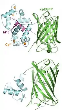
Indicators have been designed to measure ion concentrations, membrane potential, neurotransmitters, and various intracellular signaling molecules. The following list provides only examples for each class; many more have been published.
- Genetically encoded calcium indicators (GECI): A large class of tools, based on natural calcium binding proteins (calmodulin, troponin). Different affinities, kinetics, and colors (green, red) available. Read-out via fluorescence intensity (single wavelength indicators), FRET or BRET. Have been targeted to various organelles. Current version: JGCaMP8[11]
- Genetically encoded chloride indicators: Clomeleon[12]
- Genetically encoded indicators for intracellular pH (GEPhI): CypHer[13]
- Genetically encoded voltage indicators (GEVI): ArcLight[14]
- Genetically encoded vesicle fusion sensors: Synapto-pHluorin, Synaptophysin-pHluorin[15]
- Genetically encoded glutamate sensors: GluSnFR[16]
- Genetically encoded GABA sensors: iGABASnFR[17]
- Genetically encoded dopamine sensors: dLight1,[18] GRAB-DA[19]
- Genetically encoded serotonin sensors: GRAB5-HT[20]
- Genetically encoded norepinephrine sensors: GRABNE[21]
- Genetically encoded ATP sensors: QUEEN-37C[22]
- Genetically encoded cAMP sensors: EPAC[23]
- Genetically encoded kinase activity sensors: CaMui,[24] SmURFP
- Genetically encoded Small G-protein sensors: FRas[25]
- Genetically encoded sensor for endocannabinoid activity: GRABeCB2.0[26]
References
- Miyawaki A, Llopis J, Heim R, McCaffery JM, Adams JA, Ikura M, Tsien RY (August 1997). "Fluorescent indicators for Ca2+ based on green fluorescent proteins and calmodulin". Nature. 388 (6645): 882–7. Bibcode:1997Natur.388..882M. doi:10.1038/42264. PMID 9278050. S2CID 13745050.
- Kerr R, Lev-Ram V, Baird G, Vincent P, Tsien RY, Schafer WR (June 2000). "Optical imaging of calcium transients in neurons and pharyngeal muscle of C. elegans". Neuron. 26 (3): 583–94. doi:10.1016/s0896-6273(00)81196-4. PMID 10896155. S2CID 311998.
- Fiala A, Spall T, Diegelmann S, Eisermann B, Sachse S, Devaud JM, et al. (October 2002). "Genetically expressed cameleon in Drosophila melanogaster is used to visualize olfactory information in projection neurons". Current Biology. 12 (21): 1877–84. doi:10.1016/s0960-9822(02)01239-3. PMID 12419190. S2CID 6312049.
- Higashijima S, Masino MA, Mandel G, Fetcho JR (December 2003). "Imaging neuronal activity during zebrafish behavior with a genetically encoded calcium indicator". Journal of Neurophysiology. 90 (6): 3986–97. doi:10.1152/jn.00576.2003. PMID 12930818. S2CID 2230173.
- Ji G, Feldman ME, Deng KY, Greene KS, Wilson J, Lee JC, et al. (May 2004). "Ca2+-sensing transgenic mice: postsynaptic signaling in smooth muscle". The Journal of Biological Chemistry. 279 (20): 21461–8. doi:10.1074/jbc.M401084200. PMID 14990564.
- Nakai J, Ohkura M, Imoto K (February 2001). "A high signal-to-noise Ca2+ probe composed of a single green fluorescent protein". Nature Biotechnology. 19 (2): 137–41. doi:10.1038/84397. PMID 11175727. S2CID 30254550.
- Chen TW, Wardill TJ, Sun Y, Pulver SR, Renninger SL, Baohan A, et al. (July 2013). "Ultrasensitive fluorescent proteins for imaging neuronal activity". Nature. 499 (7458): 295–300. Bibcode:2013Natur.499..295C. doi:10.1038/nature12354. PMC 3777791. PMID 23868258.
- Ravotto, Luca; Duffet, Loïc; Zhou, Xuehan; Weber, Bruno; Patriarchi, Tommaso (2020). "A Bright and Colorful Future for G-Protein Coupled Receptor Sensors". Frontiers in Cellular Neuroscience. 14: 67. doi:10.3389/fncel.2020.00067. ISSN 1662-5102. PMC 7098945. PMID 32265667.
- Jones-Tabah, Jace; Mohammad, Hanan; Hadj-Youssef, Shadi; Kim, Lucy E. H.; Martin, Ryan D.; Benaliouad, Faïza; Tanny, Jason C.; Clarke, Paul B. S.; Hébert, Terence E. (2020-09-02). "Dopamine D1 receptor signalling in dyskinetic Parkinsonian rats revealed by fiber photometry using FRET-based biosensors". Scientific Reports. 10 (1): 14426. doi:10.1038/s41598-020-71121-8. ISSN 2045-2322. PMC 7468292. PMID 32879346.
- Verfasser, Sofroniew, Nicholas James. Q & A: The brain under a mesoscope: the forest and the trees. OCLC 1188624635.
- Zhang, Yan; Rózsa, Márton; Bushey, Daniel; Jihong Zheng; Reep, Daniel; Yajie Liang; Broussard, Gerard Joey; Tsang, Arthur; Getahun Tsegaye; Patel, Ronak; Sujatha Narayan (2020). "jGCaMP8 Fast Genetically Encoded Calcium Indicators": 361685 Bytes. doi:10.25378/JANELIA.13148243. Cite journal requires
|journal=(help) - Berglund, Ken; Schleich, Wolfram; Wang, Hong; Feng, Guoping; Hall, William C.; Kuner, Thomas; Augustine, George J. (2008). "Imaging synaptic inhibition throughout the brain via genetically targeted Clomeleon". Brain Cell Biology. 36 (1–4): 101–118. doi:10.1007/s11068-008-9031-x. ISSN 1559-7105. PMC 2674236. PMID 18850274.
- Han, Junyan; Burgess, Kevin (2009-10-16). "Fluorescent Indicators for Intracellular pH". Chemical Reviews. 110 (5): 2709–2728. doi:10.1021/cr900249z. ISSN 0009-2665.
- Jin, Lei; Han, Zhou; Platisa, Jelena; Wooltorton, Julian R.A.; Cohen, Lawrence B.; Pieribone, Vincent A. (2012). "Single Action Potentials and Subthreshold Electrical Events Imaged in Neurons with a Fluorescent Protein Voltage Probe". Neuron. 75 (5): 779–785. doi:10.1016/j.neuron.2012.06.040. PMC 3439164. PMID 22958819.
- Granseth, Björn; Odermatt, Benjamin; Royle, Stephen J.; Lagnado, Leon (2006). "Clathrin-Mediated Endocytosis Is the Dominant Mechanism of Vesicle Retrieval at Hippocampal Synapses". Neuron. 51 (6): 773–786. doi:10.1016/j.neuron.2006.08.029.
- Marvin, Jonathan S.; Scholl, Benjamin; Wilson, Daniel E.; Podgorski, Kaspar; Kazemipour, Abbas; Müller, Johannes Alexander; Schoch, Susanne; Quiroz, Francisco José Urra; Rebola, Nelson; Bao, Huan; Little, Justin P. (2018). "Stability, affinity, and chromatic variants of the glutamate sensor iGluSnFR". Nature Methods. 15 (11): 936–939. doi:10.1038/s41592-018-0171-3. ISSN 1548-7105. PMC 6394230. PMID 30377363.
- Marvin, Jonathan S.; Shimoda, Yoshiteru; Magloire, Vincent; Leite, Marco; Kawashima, Takashi; Jensen, Thomas P.; Kolb, Ilya; Knott, Erika L.; Novak, Ondrej; Podgorski, Kaspar; Leidenheimer, Nancy J. (2019). "A genetically encoded fluorescent sensor for in vivo imaging of GABA". Nature Methods. 16 (8): 763–770. doi:10.1038/s41592-019-0471-2. ISSN 1548-7105.
- Patriarchi, Tommaso; Cho, Jounhong Ryan; Merten, Katharina; Howe, Mark W.; Marley, Aaron; Xiong, Wei-Hong; Folk, Robert W.; Broussard, Gerard Joey; Liang, Ruqiang; Jang, Min Jee; Zhong, Haining (2018-06-29). "Ultrafast neuronal imaging of dopamine dynamics with designed genetically encoded sensors". Science. 360 (6396): eaat4422. doi:10.1126/science.aat4422. ISSN 0036-8075. PMC 6287765. PMID 29853555.
- Labouesse, Marie A.; Cola, Reto B.; Patriarchi, Tommaso (2020-10-28). "GPCR-Based Dopamine Sensors—A Detailed Guide to Inform Sensor Choice for In Vivo Imaging". International Journal of Molecular Sciences. 21 (21): 8048. doi:10.3390/ijms21218048. ISSN 1422-0067. PMC 7672611. PMID 33126757.
- Wan, Jinxia; Peng, Wanling; Li, Xuelin; Qian, Tongrui; Song, Kun; Zeng, Jianzhi; Deng, Fei; Hao, Suyu; Feng, Jiesi; Zhang, Peng; Zhang, Yajun (2021). "A genetically encoded sensor for measuring serotonin dynamics". Nature Neuroscience. 24 (5): 746–752. doi:10.1038/s41593-021-00823-7. ISSN 1546-1726. PMC 8544647. PMID 33821000.
- Feng, Jiesi; Zhang, Changmei; Lischinsky, Julieta E.; Jing, Miao; Zhou, Jingheng; Wang, Huan; Zhang, Yajun; Dong, Ao; Wu, Zhaofa; Wu, Hao; Chen, Weiyu (May 2019). "A Genetically Encoded Fluorescent Sensor for Rapid and Specific In Vivo Detection of Norepinephrine". Neuron. 102 (4): 745–761.e8. doi:10.1016/j.neuron.2019.02.037. PMC 6533151. PMID 30922875.
- Yaginuma, Hideyuki; Okada, Yasushi (2021-10-09). "Live cell imaging of metabolic heterogeneity by quantitative fluorescent ATP indicator protein, QUEEN-37C": 2021.10.08.463131. doi:10.1101/2021.10.08.463131. Cite journal requires
|journal=(help) - Klarenbeek, Jeffrey; Goedhart, Joachim; Batenburg, Aernoud van; Groenewald, Daniella; Jalink, Kees (2015-04-14). "Fourth-Generation Epac-Based FRET Sensors for cAMP Feature Exceptional Brightness, Photostability and Dynamic Range: Characterization of Dedicated Sensors for FLIM, for Ratiometry and with High Affinity". PLOS One. 10 (4): e0122513. doi:10.1371/journal.pone.0122513. ISSN 1932-6203. PMC 4397040. PMID 25875503.
- Lee, Seok-Jin R.; Escobedo-Lozoya, Yasmin; Szatmari, Erzsebet M.; Yasuda, Ryohei (2009). "Activation of CaMKII in single dendritic spines during long-term potentiation". Nature. 458 (7236): 299–304. doi:10.1038/nature07842. ISSN 0028-0836. PMC 2719773. PMID 19295602.
- Oliveira, Ana F.; Yasuda, Ryohei (2013-01-14). "An Improved Ras Sensor for Highly Sensitive and Quantitative FRET-FLIM Imaging". PLOS One. 8 (1): e52874. doi:10.1371/journal.pone.0052874. ISSN 1932-6203. PMC 3544822. PMID 23349692.
- Dong, Ao; He, Kaikai; Dudok, Barna; Farrell, Jordan S.; Guan, Wuqiang; Liput, Daniel J.; Puhl, Henry L.; Cai, Ruyi; Wang, Huan; Duan, Jiali; Albarran, Eddy (2021-11-11). "A fluorescent sensor for spatiotemporally resolved imaging of endocannabinoid dynamics in vivo". Nature Biotechnology: 1–12. doi:10.1038/s41587-021-01074-4. ISSN 1546-1696.
| Optogenetic actuators | |
|---|---|
| Optogenetic sensors | |
| Related techniques | |
